A case of trigeminal peripheral nerve sheath tumour in a dog diagnosed with tru-cut biopsy
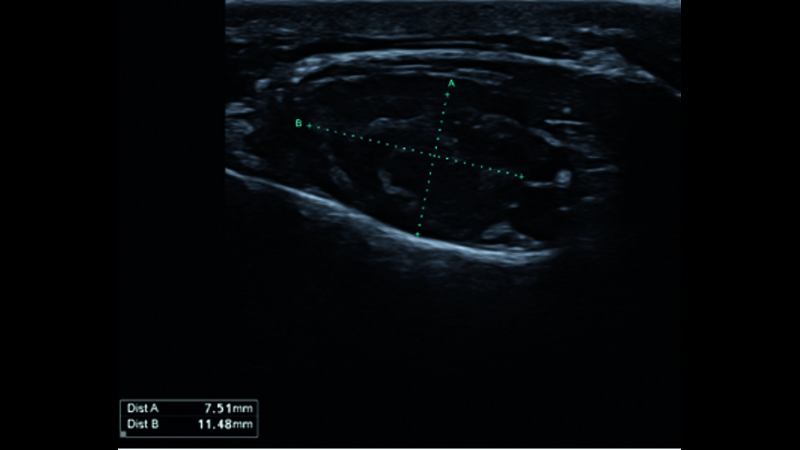
Peripheral nerve sheath tumours present a diagnostic challenge. Only one case where ante-mortem diagnosis was achieved following a wedge biopsy is described in the literature; this is the first case described of canine peripheral nerve sheath tumour diagnosed via tru-cut biopsy. This technique represents a less invasive approach to diagnose trigeminal peripheral nerve sheath tumours. A 7-year-old female Bull Terrier presented with an 8-week history of left temporalis muscle atrophy and facial irritation. A subcutaneous mass was detected on the left maxilla at the level of the infraorbital foramen. Exophthalmos, atrophy of the temporalis and masseter muscles, and reduced sensation of the nostril were detected on the left side. Magnetic resonance imaging revealed enlargement and hyperintense signal intensity compared to muscle signal intensity in T2-weighted magnetic resonance images of the left mandibular and maxillary branches of the trigeminal nerve, extending into the infraorbital branch of the maxillary nerve, and enlargement of the infraorbital foramen was noted. Affected nerves showed marked heterogeneous contrast enhancement. Six ultrasound-guided tru-cut biopsies of the trigeminal nerve were taken, and histopathology and immunohistochemistry confirmed the diagnosis of a peripheral nerve sheath tumour. Surgical excision and radiation therapy were discussed, but a palliative approach was elected, and gabapentin was prescribed. The facial irritation improved but did not resolve. The patient was lost to follow up after a few weeks.
Valentina Granziera -
Neringa Alisauskaite -